Home
/ Posterior View Of Heart Anatomy - Functional Anatomy Of The Cardiovascular System Clinical Gate : Intraoperatively, the anatomy of the heart is viewed from the right side of the supine patient via a median sternotomy incision.
Posterior View Of Heart Anatomy - Functional Anatomy Of The Cardiovascular System Clinical Gate : Intraoperatively, the anatomy of the heart is viewed from the right side of the supine patient via a median sternotomy incision.
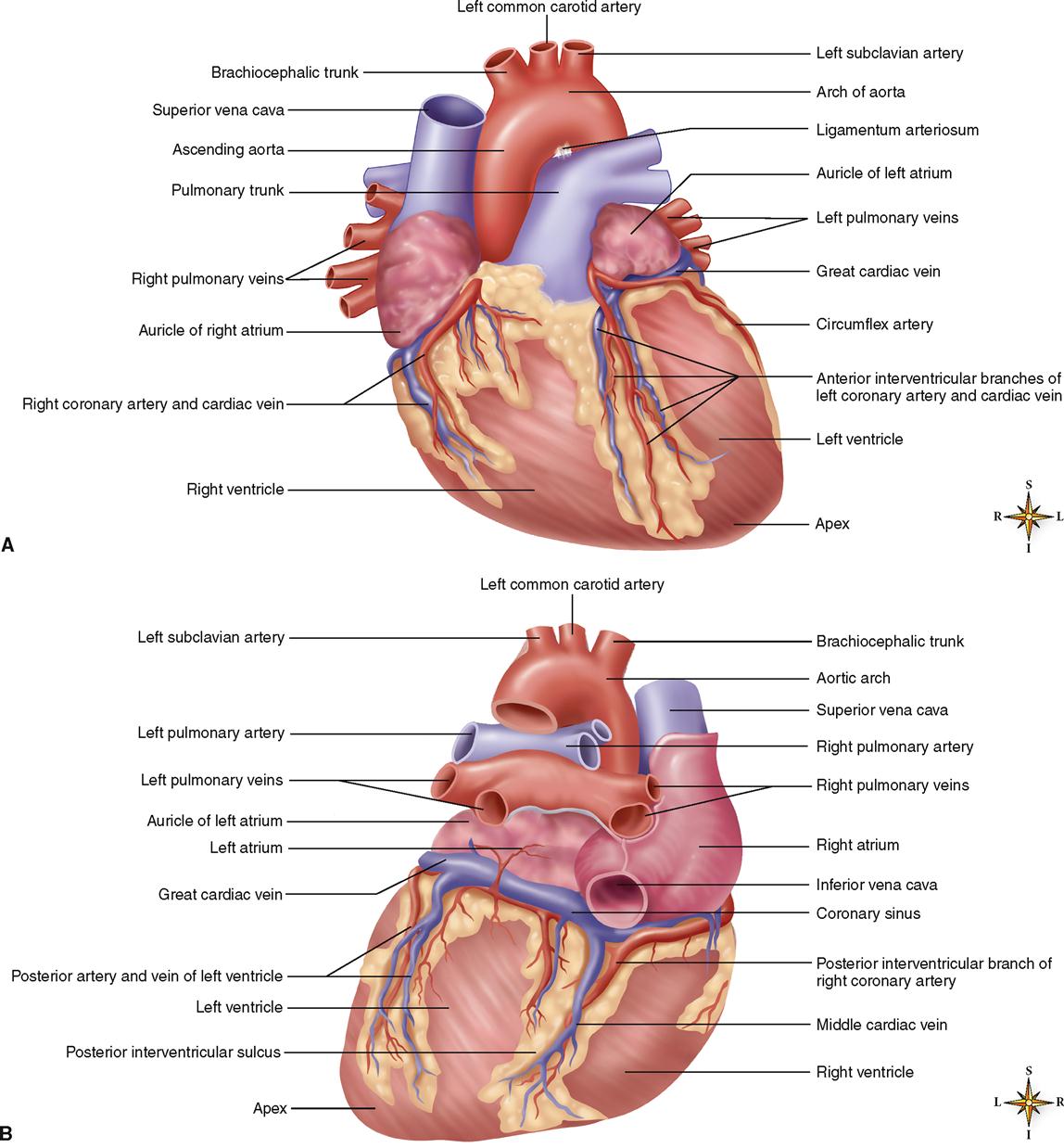
Posterior View Of Heart Anatomy - Functional Anatomy Of The Cardiovascular System Clinical Gate : Intraoperatively, the anatomy of the heart is viewed from the right side of the supine patient via a median sternotomy incision.. This is an online quiz called heart anatomy posterior view. There is a printable worksheet available for download here so you can take the quiz with pen and paper. This image shows the anatomy of the heart from external posterior view showing the different parts and features of the heart with the related vessels showing: Posterior view of heart anatomy in this image you will find superior vena cava right pulmonary artery right pulmonary veins right atrium inferior vena cava coronary sinus right coronary artery coronary sulcus posterior interventricular artery in it. On its superior end, the base of the heart is attached to the aorta,continue scrolling to read more below.
There is a printable worksheet available for download here so you can take the quiz with pen and paper. Posterior view of heart anatomy in this image, you will find superior vena cava, right pulmonary artery, right pulmonary veins, right atrium, inferior vena cava, coronary sinus, right coronary artery, coronary sulcus, posterior interventricular artery in it. This image shows the anatomy of the heart from external posterior view showing the different parts and features of the heart with the related vessels showing: On its superior end, the base of the heart is attached to the aorta,continue scrolling to read more below. With the buttons on the right you can light up different anatomic structures.
4 Posterior View Of The Human Heart Download Scientific Diagram from www.researchgate.net This image shows the anatomy of the heart from external posterior view showing the different parts and features of the heart with the related vessels showing: The heart anatomy * chapter 18, cardiovascular system * congestive heart failure (chf) congestive heart failure (chf) is caused by: Drawings surface anatomy of the heart drawings of the surface anatomy of the normal heart, anterior and posterior, with english labels. Normally, the arteries run through grooves of the epicardial surface of the heart. This image shows the anatomy of the heart from external posterior view showing the different parts and features of the heart with the related vessels showing: Posterior view of heart anatomy in this image, you will find superior vena cava, right pulmonary artery, right pulmonary veins, right atrium, inferior vena cava, coronary sinus, right coronary artery, coronary sulcus, posterior interventricular artery in it. Start studying heart anatomy posterior view. The regions of the body are labeled in boldface.
Intraoperatively, the anatomy of the heart is viewed from the right side of the supine patient via a median sternotomy incision.
There is a printable worksheet available for download here so you can take the quiz with pen and paper. This image shows the anatomy of the heart from external posterior view showing the different parts and features of the heart with the related vessels showing: Contribute by gray's anatomy plates A photo of the posterior view of a plastinated normal heart, where highlights on structures can be switched on and off, by univ. Download 1,489 posterior view body stock illustrations, vectors & clipart for free or amazingly low rates! In this figure, an posterior view of the heart is visible with opened atria, so the anatomy of the ventral wall can be seen. Overview of the external anatomy of the heart, posterior view reset help right ventricle left atnum abrtic arch left ventricle left pulmonary veins inferior vena cava leit pulmonary artery circumflex artery right coronary artery posterior interventricular artery superior vena cava right atrium. Start studying heart anatomy posterior view. Just pick an audience or yourself and itll end up in. The heart is located in the thoracic cavity medial to the lungs and posterior to the sternum. Posterior view of heart anatomy in this image, you will find superior vena cava, right pulmonary artery, right pulmonary veins, right atrium, inferior vena cava, coronary sinus, right coronary artery, coronary sulcus, posterior interventricular artery in it. The anterior interventricular sulcus is visible on the anterior surface of the heart, whereas the posterior interventricular sulcus is visible on the posterior surface of the heart. Position lies within the pericardium in middle mediastinum behind the body of sternum and the 2nd to 6th costal cartilages in front of the 5th to 8th thoracic vertebrae a third of it lies to the right of median plane and 2/3 to the left anterior to the vertebral column, posterior.
Download 1,489 posterior view body stock illustrations, vectors & clipart for free or amazingly low rates! Overview of the external anatomy of the heart, posterior view reset help right ventricle left atnum abrtic arch left ventricle left pulmonary veins inferior vena cava leit pulmonary artery circumflex artery right coronary artery posterior interventricular artery superior vena cava right atrium. Located between the left and right ventricles are two additional sulci that are not as deep as the coronary sulcus. The anterior interventricular sulcus is visible on the anterior surface of the heart, whereas the posterior interventricular sulcus is visible on the posterior surface of the heart. Anatomy of the human heart posterior view.
Anatomy Of The Cardiovascular System Basicmedical Key from basicmedicalkey.com They are named the left and right coronary arteries, and arise from the left and right aortic sinuses within the aorta. Surface anatomy and features of the heart and the pericardium the pericardium: Its main function is to prevent sudden overfilling of the heart. With the buttons on the right you can light up different anatomic structures. Definition / description (fig 1): This image shows the anatomy of the heart from external posterior view showing the different parts and features of the heart with the related vessels showing: Intraoperatively, the anatomy of the heart is viewed from the right side of the supine patient via a median sternotomy incision. The heart is shaped as a quadrangular pyramid, and orientated as if the pyramid has fallen onto one of its sides so that its base faces the posterior thoracic wall, and its apex is pointed toward the anterior thoracic wall.
The heart is shaped as a quadrangular pyramid, and orientated as if the pyramid has fallen onto one of its sides so that its base faces the posterior thoracic wall, and its apex is pointed toward the anterior thoracic wall.
Surface anatomy and features of the heart and the pericardium the pericardium: Drawings surface anatomy of the heart drawings of the surface anatomy of the normal heart, anterior and posterior, with english labels. Posterior view of heart anatomy in this image you will find superior vena cava right pulmonary artery right pulmonary veins right atrium inferior vena cava coronary sinus right coronary artery coronary sulcus posterior interventricular artery in it. Learn vocabulary, terms, and more with flashcards, games, and other study tools. The heart is located in the thoracic cavity medial to the lungs and posterior to the sternum. Download 1,489 posterior view body stock illustrations, vectors & clipart for free or amazingly low rates! There is a printable worksheet available for download here so you can take the quiz with pen and paper. The anterior interventricular sulcus is visible on the anterior surface of the heart, whereas the posterior interventricular sulcus is visible on the posterior surface of the heart. Start studying heart anatomy posterior view. Heart anatomy in detail anterior view. Lecture on anatomy of the heart ( drnnamanisamuel@gmail.com) 3. This is an online quiz called heart anatomy posterior view. The heart anatomy * chapter 18, cardiovascular system * congestive heart failure (chf) congestive heart failure (chf) is caused by:
This image shows the anatomy of the heart from external posterior view showing the different parts and features of the heart with the related vessels showing: The structures initially seen from this perspective include the superior vena cava, right atrium, right ventricle, pulmonary artery, and aorta. Just pick an audience or yourself and itll end up in. Normally, the arteries run through grooves of the epicardial surface of the heart. There is a printable worksheet available for download here so you can take the quiz with pen and paper.
View Of Human Heart Png Download Anatomical Heart Posterior View Transparent Png Vhv from www.vhv.rs In this image, you will find heart anatomy in detail anterior view, brachiocephalic view, superior vena cava, right pulmonary artery, ascending aorta, pulmonary trunk in it. Heart anatomy in detail anterior view. Intraoperatively, the anatomy of the heart is viewed from the right side of the supine patient via a median sternotomy incision. Start studying heart anatomy posterior view. With the buttons on the right you can light up different anatomic structures. The structures initially seen from this perspective include the superior vena cava, right atrium, right ventricle, pulmonary artery, and aorta. Contribute by gray's anatomy plates Start studying external heart anatomy posterior view.
The regions of the body are labeled in boldface.
With the buttons on the right you can light up different anatomic structures. Surface anatomy and features of the heart and the pericardium the pericardium: The heart is located in the thoracic cavity medial to the lungs and posterior to the sternum. In this figure, an posterior view of the heart is visible with opened atria, so the anatomy of the ventral wall can be seen. This image shows the anatomy of the heart from external posterior view showing the different parts and features of the heart with the related vessels showing: Ramus circumflexus (arteria coronaria sinistra) aorta. In addition, codominance (balanced distribution) of the arteries (the posterior descending branch comes from the rca, and the posterior left ventricular branch derives from the lcx artery) is found in less than 20% of cases. Download 1,489 posterior view body stock illustrations, vectors & clipart for free or amazingly low rates! Anatomy of the human heart posterior view. The regions of the body are labeled in boldface. The structures initially seen from this perspective include the superior vena cava, right atrium, right ventricle, pulmonary artery, and aorta. Overview of the external anatomy of the heart, posterior view reset help right ventricle left atnum abrtic arch left ventricle left pulmonary veins inferior vena cava leit pulmonary artery circumflex artery right coronary artery posterior interventricular artery superior vena cava right atrium. Contribute by gray's anatomy plates

